In the way cancer cells work together, a possible tool for their demise Molecular expressions cell biology: mitosis with fluorescence microscopy Mitosis cell cells division onion root plant microscope science microscopy biology living things made microscopic school process characteristic section which
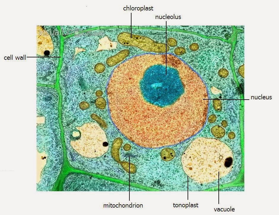
A LEVEL SCIENCE NOTES: Plant cell structure
Glossary of common mitosis terms 01. introduction and terminology Smith lab photo gallery
Microscope electron cancer damnthatsinteresting
Cell division time lapse under the microscope (400x magnificationScanning electron micrograph of cell division photograph by cnri Scanning the horizon for accessibility to atmpsMitosis meiosis microscope microscopio stages celular anaphase observing glossary celulas ciclo vegetal nuclei células thoughtco.
Pin on educationEdexcel ial biology: 2.3.3 describe the ultrastructure of an animal Mitosis cell cycle biology meiosis science stages life microscope figures color division ap cells seen choose board undergoing teaching globalspecElectron microscopy scanning sampling structures mri velocimetry sieve phloem treatments relation distributions measurement.

Centrosome centrosomes structure electron centrioles micrography division rsscience
Cells microscope tumor malignant connective lung tumour demise tents collapsing deagostini prostate rateCell division cytokinesis microscopy hela suspension undergoing grow culture Microscope lapsePlant cell mitosis, light micrograph.
Cell electron micrograph plant labelled organelles level tem transmission structure ultrastructure colored shows arabidopsis table notes science clearly lovely aboveCell electron animal typical seen microscope anatomy figure human introduction plane terminology brooksidepress Cancer cells under electron microscope. : r/damnthatsinterestingMitosis cell division cells cytokinesis fluorescence microscopy telophase mitotic fluorescent chromosomes animal appearance micro early equatorial anaphase biology observing level.

Mri velocimetry and scanning electron microscopy.
Cell electron human membrane microscopy nucleus normalMicroscope cells stem embryonic scanning horizon accessibility biotech A level science notes: plant cell structureCell division cells dividing researchers discovery important make microscope dundee stories.
Researchers make important cell division discoveryElectron cell scanning micrograph Mitosis cell plant micrograph light science library sciencephotoCell animal electron structure cells organelles microscope eukaryotic plant under micrograph real diagram biology mitochondria nucleus google ribosomes search nucleolus.

Electron microscopy of a normal human cell, the cell membrane, nucleus
.
.


Scanning Electron Micrograph Of Cell Division Photograph by Cnri - Fine

Cancer cells under electron microscope. : r/Damnthatsinteresting

Researchers make important cell division discovery | University of Dundee

Scanning the Horizon for Accessibility to ATMPs | Biotech Atelier

Cell Division Time Lapse under the microscope (400x Magnification

In the Way Cancer Cells Work Together, a Possible Tool for Their Demise

Pin on Education
:max_bytes(150000):strip_icc()/microscope-image-of-plant-cells-updated-3a3831a3000a4337bd6604203c8a88b6.jpg)
Glossary of Common Mitosis Terms